Diagnosis is a Multistage Process

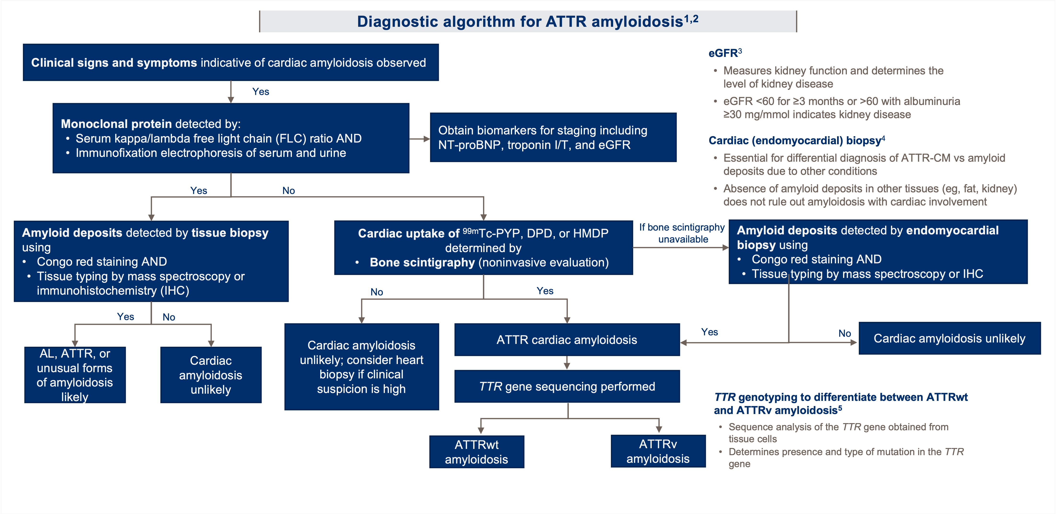
Diagnosis of amyloidosis involves a combination of blood, urine, imaging, and biopsy tests, which are mainly done in sequence, or in parallel [1]
Responsive Image
Some tests are done only once to establish or confirm a diagnosis, whereas others are done multiple times to diagnose, monitor disease progression, and determine response to treatment
Blood tests
  • TnT (cardiomyopathy)
  • NT-proBNP (cardiomyopathy)
  • dFLC (plasma cell dyscrasia)
  • ALP (liver involvement)
  • Bilirubin (liver involvement)
  • eGFR (kidney damage)
Imaging tests
  • Presence of amyloid deposits in different organs (e.g., CMR/PET imaging tracers)
  • Extent of amyloid deposits in organs (heart)
  • Extent of damage to organs (heart)
Biopsy tests
  • Visual confirmation of amyloid deposits in organs
  • Typing of the amyloid precursor protein
Urine tests
  • Proteinuria (kidney involvement)
  • Albuminuria (kidney involvement)
Other tests
  • Nerve conduction studies
  • Neuro-vegetative tests
Notes: ALP = alkaline phosphatase; eGFR = estimated glomerular filtration rate; dFLC = difference between uninvolved and involved free light chain; NT-proBNP = N-terminal pro-brain natriuretic peptide; PET = positron emission tomography; TnT = troponin T
  1. ALXN: 32_2021 Amyloidosis March KOL Perspectives_Cristina Quarta_FINAL. Citing:Amyloidosis Foundation. AL Amyloidosis. http://amyloidosis.org/facts/al/#diagnosis. Accessed September 17, 2020.

Amyloidosis Clinical Presentation & Typical Lab Findings

Responsive Image

Cardiac & Extracardiac Red Flags for CA

CA typically appears within a constellation of extracardiac signs and symptoms that are extremely useful to suspect the disease in the presence of compatible cardiac imaging findings
Cardiac and Extracardiac Amyloidosis Red Flags
Responsive Image
  • Signs and symptoms include proteinuria (even mild), macroglossia, skin bruises, and carpal tunnel syndrome, among others
  • There are also various red flags at the cardiac level, such as HF (including disproportionately high NT-proBNP) that appears to be in disproportion to ‘objective’ findings on ECG, ‘unexplained’ right HF in the presence of ostensibly ‘normal’ ventricular and valvular function, or ‘idiopathic’ pericardial effusion
  • Persistent troponin elevation, disproportionally low QRS voltage, or early conduction system disease are also signs that could evoke CA [1]
Screening for CA
Responsive Image
Responsive Image
Notes: AA = serum amyloid A amyloidosis; AApoAI = apolipoprotein AI amyloidosis; AApoAII = apolipoprotein AII amyloidosis; AApoAIV = apolipoprotein A-IV amyloidosis; Ab2M = b2-microglobulin amyloidosis; Afib = fibrinogen amyloidosis; Agel = gelsolin amyloidosis; AL = light-chain amyloidosis; ATTRv = hereditary transthyretin amyloidosis; ATTRwt = wild-type transthyretin amyloidosis; AV = atrio-ventricular; CA = cardiac amyloidosis; ECG = electrocardiogram; HF = heart failure; LV = left ventricular; NT-proBNP = N-terminal proB-type natriuretic peptide
  1. Garcia-Pavia P, et al. Diagnosis and Treatment of Cardiac Amyloidosis: A Position Statement of the ESC Working Group on Myocardial and Pericardial Diseases. Eur Heart J. 2021;42(16):1554-1568. DOI: https://doi.org/10.1093/eurheartj/ehab072.

Imaging Techniques for Suspicion of ATTR-CM

While none of these tests are typically used to confirm an ATTR-CM diagnosis, they can help doctors learn more about the patient’s heart and determine the need for additional diagnostic testing
Responsive Image
Electrocardiogram
  • Evaluates electrical conduction; helps confirm disease and determine level of cardiac involvement
  • Changes from normal pattern indicate heart stress and/or CM; the nature of the change helps identify specific cardiac dysfunction
  • Common changes include low voltage on limb leads and pseudo‐infarct pattern, atrial arrhythmia and atrioventricular block, ischemia/pseudo-necrosis patterns, conduction delays [1-3]
  • CAUTION: low voltage seen in <50% of cases with ATTR-CM5
Responsive Image
Echocardiogram (heart ultrasound) [1,2]
  • Provides comprehensive morphological and functional assessment of the heart 
  • Findings indicate generalized CM and are not specific to ATTR amyloidosis and should therefore be interpreted in the context of other evaluations
  • ATTR amyloidosis-driven CM presents with “classic” features on an echo only in advanced disease [1,2]
*Thickened walls, leading to a stiff heart and poor pumping function [4]
Responsive Image
CMR imaging [1,2]
  • Provides detailed information on the type and severity of CM
  • Measures thickness and volumes of both ventricles more accurately than an echocardiogram
  • Can assess biatrial dilatation, interatrial septal thickness, and the presence of pericardial effusion and can evaluate left ventricular filling
  • Can quantify the amyloid burden and track changes of amyloid over time through the extracellular volume 
ALXN: Acoramidis ​for the Treatment of ATTR-CM: Training Deck. November 22, 2021. 34_Acoradmidis Training Deck_22Nov21 FINAL. Citing:
  1. Firkle M, et al. Cor et Vasa. 2013;55:e60-e75.
  2. Kitaoka H, et al. Circ J. 2020;84:1610-1671.
  3. Healio. Introduction to ECG. https://www.healio.com/cardiology/learn-the-heart/ecg-review/ecg-interpretation-tutorial/introduction-to-the-ecg. Accessed August 23, 2021.
  4. Quarta CC, et al. Circulation. 2012;126:e178-e182. 
  5. Ruberg FL, Grogan M, Hanna M, Kelly JW, Maurer MS. Transthyretin Amyloid Cardiomyopathy: JACC State-of-the-Art Review. J Am Coll Cardiol. 2019;73(22):2872-91.

Potential Gender Differences in Biopsy Predictiveness

A recent study on ATTRwt-CA showed that female patients had a higher sensitivity to TTR deposition from abdominal fat than male patients [1]
  • Based on results of previous studies showing that male patients were more prone to cardiac ATTR deposition than female patients, it was speculated that females may develop cardiomyopathy at later stages of the disease, thus allowing for more amyloid accumulation in non-cardiac tissues
  • It has not been determined whether this rule applies to patients with ATTRwt, however, 80% of female patients in the cohort in whom an abdominal fat pad biopsy was performed were positive for amyloid (8 out of 10 patients)*
  • Abdominal fat biopsy is clearly less invasive than EMB, and it is suggested that abdominal fat biopsy should be performed particularly in elderly female patients with suspected ATTRwt
  • The rate of TTR detection in abdominal fat tissue was significantly higher in female than male patients
  • Conversely, all patients, both female and male, in whom EMB was performed were positive for amyloid
Results of Organ Biopsies According to Sex
Responsive Image
The Japanese female elderly patients had a small BSA, so concentric LV hypertrophy and a low-flow state may easily appear; consequently, Japanese female ATTRwt patients may be more symptomatic under conditions of HFpEF even before the advanced phase of ATTRwt, with reduced LVEF and moderately increased LV wall thickness. Thus, clinicians should be aware of the possibility of ATTRwt in female patients with a low threshold even if they are at the stage of preserved EF and mild LV hypertrophy; use of lower cut-off values or cardiac dimensions indexed by BSA to diagnose females has been discussed because they are less likely to meet the diagnostic wall thickness threshold. [1]
Notes: BSA = body surface area; EMB = endomyocardial biopsy; LV = left ventricular; LVEF = LV ejection fraction

Notes: * Although the number of patients was relatively small, female ATTRwt patients had a higher sensitivity for TTR detection in abdominal fat tissue, smaller LV cavity, milder LVH, and a higher frequency of preserved LVEF than male ATTRwt patients; there have been few studies in which sex differences in ATTRwt have been investigated, and information on japanese female patients with ATTRwt remains insufficient

  1. Ochi Y, Kubo T, Baba Y, et al. Wild-Type Transthyretin Amyloidosis in Female Patients - Consideration of Sex Differences. Circ Rep. 2021;3(8):465-471. Published 2021 Jun 29. doi:10.1253/circrep.CR-21-0067.

Diagnostic Algorithm: Japanese Circulation Society (JCS) 2020 Guidelines

japan Responsive Image
Responsive Image
Glossary:
 
  • Macroglossia, enlargement of the tongue​
  • Magnetic resonance imaging (MRI), a medical imaging technique that uses strong magnetic fields and radio waves to produce detailed images of the inside of the body​
  • Myeloma protein (M protein), an abnormal antibody or fragment that is excessively produced by the monoclonal proliferation of B lymphocytes and plasma cells. M protein typically occurs in disorders known as monoclonal gammopathies, paraproteinemias, or plasma cell dyscrasias such as multiple myeloma
  1. ALXN: Acoramidis ​for the Treatment of ATTR-CM: Training Deck. November 22, 2021. 34_Acoradmidis Training Deck_22Nov21 FINAL. Citing: Kitaoka H, et al. Circ J. 2020;84:1610-1671.

Diagnostic Algorithm for ATTR Amyloidosis

usa Responsive Image
Responsive Image
Glossary:
 
  • ⁹⁹ᵐ technetium (Tc)-3,3-diphosphono-1,2-propanodicarboxylic acid (DPD), ⁹⁹ᵐTc-hydroxymethylene diphosphonate (HMDP), ⁹⁹ᵐTc-pyrophosphate (PYP), tracers used in bone scintigraphy​
  • Electrophoresis, a laboratory technique to separate and analyze macromolecules such as DNA, RNA, and proteins based on their size and electrical charge​
  • Endomyocardial, relating to the innermost layer of the heart that lines the chambers (endocardium) and the middle muscular layer of the heart (myocardium)​
  • Free light chain (FLC), light chain not bound to a heavy chain to form immunoglobulin (antibody)​
  • Immunofixation, a test for detecting the presence and type of certain proteins (monoclonal antibodies, immunoglobulins)​
  • Immunohistochemistry (IHC), a laboratory technique that allows imaging of different components in tissues by using fluorescently-tagged or appropriately-labeled antibodies that bind specifically to target antigens (markers)​
  • Kappa (κ) subtype, a subtype of immunoglobulin light chain; twice as common as λ in humans ​
  • Lambda (λ) subtype, a subtype of immunoglobulin light chain; half as common as κ in humans​
  • Mass spectroscopy, an analytical technique that identifies molecules by fragmenting them into charged species (ions) and measuring their mass-to-charge ratios​
  • Scintigraphy, a diagnostic technique in which a scintillation counter or similar detector is used with a radioactive tracer to obtain an image of an organ or a record of its functioning​
  • Troponin T (TnT), part of a protein complex integral to contraction of heart and skeletal muscle; elevation indicates injury to the heart
  1. Ruberg FL, et al. J Am Coll Cardiol. 2019;73:2872-2891.
  2. Kittleson MM, et al. Circulation. 2020;142:e7-e22. Erratum in: Circulation. 2021;144:e11.
  3. National Kidney Foundation. Estimated glomerular filtration rate (eGFR). Accessed August 16, 2021. https://www.kidney.org/atoz/content/gfr.
  4. Veinot JP. Can J Cardiol. 2009;25(2):e55-e56.
  5. Maurer MS, et al. Circ Heart Fail. 2019;12:e006075.

Staging ATTR Amyloidosis

Two staging systems have been proposed in patients with ATTR amyloidosis (Mayo Clinic & NAC) [1]
Mayo Clinic Staging System (Grogan, et al.)
N=360 ATTRwt patients
  • The first published staging system for ATTRwt is based on serum levels of NT-proBNP and cardiac troponin T
  • Thresholds of troponin T (0.05 ng/ml) and NT-proBNP (3000 pg/ml) were used
  • The respective four-year overall survival estimates were 57%, 42%, and 18% for stage I (both values below cutoff), stage II (one above), and stage III (both above), respectively [1,2]
NOTE: The NAC study authors noted that the Mayo Clinic system “is confounded by current worldwide use of five generations of troponin assays made by various manufacturers, with different centers favoring different assays.” [3]**
Responsive Image
Responsive Image

Notes: Notes: BNP = brain natriuretic peptide; NAC = National Amyloidosis Centre (UK); NT-proBNP = N-terminal pro b-type natriuretic peptide​

Notes: * (Top left) Death rates based on troponin T cutpoint of 0.05 ng/ml. Numbers below the plot represent number of subjects at risk at each time point. The age- and sex-adjusted hazard ratio (HR) was 2.34 (95% confidence interval [CI]: 1.46 to 3.76) with a c-statistic of 0.72 (95% CI: 0.66 to 0.78). Cross-validated c-statistic was 0.71 (95% CI: 0.65 to 0.78). (Top right) Death rates based on N-terminal pro–B-type natriuretic peptide (NT-proBNP) cutpoint of 3,000 pg/ml. Numbers below the plot represent number of subjects at risk at each time point. The age- and sex-adjusted HR was 2.22 (95% CI: 1.36 to 3.60) with a c-statistic of 0.72 (95% CI: 0.65 to 0.78). Cross validated c-statistic was 0.71 (95% CI: 0.65 to 0.78). (Bottom) Death rates based on staging system, overlaid with expected survival based on age and sex. Numbers below the plot represent numbers of subjects at risk at each time point. The age- and sex-adjusted HR was 1.42 (95% CI: 0.79 to 2.57) for 1 above and 3.60 (95% CI: 2.02 to 6.42) for 2 above, with a c-statistic of 0.74 (95% CI: 0.68 to 0.80). Cross-validated c-statistic was 0.73 (95% CI: 0.67 to 0.79)2​


Notes: ** These include newer high-sensitivity troponin T (hs-TnT) assays, which not only differ in sensitivity but also give different numerical results to older assays, and troponin I assays3

Two staging systems have been proposed in patients with ATTR amyloidosis amyloidosis (Mayo Clinic & NAC) [1]
NAC Staging System (Gillmore, et al.)
N=869 ATTR-CM patients (553 with ATTRwt and 316 with ATTRv)
  • The second staging system, validated in both ATTRwt and ATTRv, is based on serum levels of NT-proBNP and eGFR
  • Stage I is defined as NT-proBNP ≤3000 ng/L and eGFR ≥45 mL/min, Stage III is defined as NT-proBNP >3000 ng/L and eGFR <45 mL/min, and the remainder were Stage II
  • Median survival among Stage I patients was 69.2 months, Stage II patients 46.7 months, and Stage III patients 24.1 months [1,3]
Kaplan–Meier curves showing survival probabilities in 553 patients with ATTRwt-CA stratified by disease stage
Responsive Image
Kaplan–Meier curves showing survival probabilities in 318 patients with ATTR-CA (validation cohort) stratified by disease stage
Responsive Image
Notes: BNP = brain natriuretic peptide; eGFR = estimated glomerular filtration rate; NAC = National Amyloidosis Centre (UK); NT-proBNP = N-terminal pro b-type natriuretic peptide
Notes: * These include newer high-sensitivity troponin T (hs-TnT) assays, which not only differ in sensitivity but also give different numerical results to older assays, and troponin I assays
  1. Fontana, Marianna. Mar. 23, 2021. Amyloid cardiomyopathy: Treatment and prognosis. UptoDate. https://www.uptodate.com/contents/amyloid-cardiomyopathy-treatment-and-prognosis#H89526001.
  2. Grogan M, Scott CG, Kyle RA, Zeldenrust SR, Gertz MA, Lin G, Klarich KW, Miller WL, Maleszewski JJ, Dispenzieri A. Natural History of Wild-Type Transthyretin Cardiac Amyloidosis and Risk Stratification Using a Novel Staging System. J Am Coll Cardiol. 2016 Sep 6;68(10):1014-20. doi: 10.1016/j.jacc.2016.06.033. Erratum in: J Am Coll Cardiol. 2017 Jun 13;69(23):2882. PMID: 27585505.
  3. Gillmore JD, Damy T, Fontana M, Hutchinson M, Lachmann HJ, Martinez-Naharro A, Quarta CC, Rezk T, Whelan CJ, Gonzalez-Lopez E, Lane T, Gilbertson JA, Rowczenio D, Petrie A, Hawkins PN. A new staging system for cardiac transthyretin amyloidosis. Eur Heart J. 2018 Aug 7;39(30):2799-2806. doi: 10.1093/eurheartj/ehx589. PMID: 29048471.