Steps to Diagnosis of ATTRwt Amyloidosis

Japan Responsive Image
Responsive Image

ATTR-CM Diagnostic Algorithm per JCS Guidelines

Responsive Image
Source: Kitaoka H, et al. JCS 2020 guideline on diagnosis and treatment of cardiac amyloidosis. Circ J. 2020. 84(9), 1610-1671. https://doi.org/10.1253/circj.CJ-20-0110; THRIVE Consulting Analysis.
  1.  ALXN: 210201_ALXN2060_Diagnostic summary

Diagnostic Criteria

japan Responsive Image
According to the current Japanese treatment guidelines, there are two diagnostic criteria: Definite and Probable; for definite diagnosis, biopsy is required
Responsive Image
Definite: A+B+C+E+G1
Probable: A+D+E+F+G2
*Reimbursement of scintigraphy to diagnose ATTR amyloidosis was approved in Japan in November 2020
  1. Kitaoka H, et al. JCS 2020 guideline on diagnosis and treatment of cardiac amyloidosis. Circ J. 2020. 84(9), 1610-1671. https://doi.org/10.1253/circj.CJ-20-0110.

Imaging Studies in Systemic Amyloidosis

Cardiac imaging has transformed the diagnostic field of ATTR amyloidosis
Responsive Image
(a) Parasternal long axis Echo demonstrating typical findings of CA with marked thickening of the IVS and PW of the LV; (b) Classic strain pattern of cardiac amyloid demonstrating a ‘bulls‐eye’ pattern of GLS*; with normal GLS the image would be all red [1]
  • Echocardiogram with strain imaging is a cornerstone in establishing heart involvement
    • Echocardiography characteristically demonstrates thickened heart walls, a restrictive diastolic filling pattern and a characteristic strain imaging pattern 
  • CMR imaging helps establish heart involvement where Echo findings are equivocal
    • The hallmark feature of CMR in CA is LGE, which indicates an expansion of the extracellular volume caused by the amyloid deposits
    • LGE was shown to have independent prognostic impact in AL and ATTR CA
Responsive Image
(a) Planar view of 99mTc‐PYP showing increased uptake over the cardiac silhouette**; (b) 99mTc‐PYP SPECT of the same patient, localizing the radioactive tracer uptake to the myocardial ventricular walls; note also a tracer uptake in bony structures, as expected [1]
  • Bonescintigraphy using technetium-labelled bisphosphonate (99mTc-DPD, 99mTc-HMDP or 99mTc-PYP) can show myocardial uptake in patients with ATTR-CA
    • Absent a monoclonal protein, a positive bone scintigraphy is highly sensitive (99%) and specific (86%) for ATTR-CA; therefore, it may replace the need for a tissue diagnosis
    • However, bone scintigraphy lacks specificity for ATTR amyloidosis in ATTR patients who have a concomitant monoclonal protein (detected in up to 19–39% of ATTRwt patients)
  • Therefore, a complete monoclonal protein evaluation that includes serum and urine electrophoresis / immunofixation and serum free light-chain assay should be performed
  • A SPECT-CT improves diagnostic accuracy over planar bone scintigraphy views, by localizing tracer uptake to the myocardium
  • PET-CT with an amyloid-targeting tracer is an emerging tool to assess amyloid disease extent
    • 18F-florbetapir PET-CT was able to detect clinically recognized and unrecognized amyloid deposits in various organs
  • Use of other imaging modalities in amyloidosis should be based on clinical suspicion and may include CT imaging of chest and abdomen to assess for lung and liver/spleen involvement, assessment of associated lymphadenopathy in cases suspected to be lymphoma-associated amyloidosis and MRI of the brain in ATTR amyloidosis if leptomeningeal involvement is suspected

 
Deep Dive
Imaging techniques for suspicion of ATTR-CM
Go to Deep Dive
Notes: CA = cardiac amyloidosis; CMR = cardiac magnetic resonance; GLS = global longitudinal peak systolic left ventricular strain; IVS = intraventricular septum; LGE = late gadolinium enhancement; LV = left ventricle; PW = posterior wall; SPECT-CT = single-photon emission computed tomography–computed tomography

Notes: * The GLS calculated to −8% compared to a normal range of −18% or higher negative number; with normal GLS the image would be all red​

Notes: ** H/CL ratio refers to the ratio between tracer retention in the heart versus the contralateral chest. Ratio greater than 1.3 (imaging 3 hours post injection; 1.5 at 1 hour) is considered positive for ATTR amyloidosis in the right clinical context. The H/CL ratio in this case is 1.7.

  1. Muchtar E, et al. J Intern Med. 2021;289:268-292.

Nuclear Imaging Techniques for Diagnosing ATTR Amyloidosis

While endocardial biopsy has been the gold standard for diagnosing ATTR-CM because of its high sensitivity and specificity, it may become less common due to advances in imaging such as ⁹⁹ᵐTc-PYP scintigraphy [3]
⁹⁹ᵐTc-PYP scintigraphy [3]
•In technetium-labeled pyrophosphate (⁹⁹ᵐTc-PYP) scintigraphy, a type of bone scintigraphy, radiolabeled pyrophosphate is injected intravenously into the patient followed by planar chest imaging for diagnosing ATTR amyloidosis [3,6]
Effectively confirms diagnosis of ATTR amyloidosis and the absence of AL amyloidosis [6]
Considered to have a diagnostic specificity and positive predictive value of 100% [3]
•Cardiac uptake (H/Cl: heart-to-contralateral ratio >1.6) considered an independent predictor of poor prognosis [3]
Responsive Image
⁹⁹ᵐTc-PYP scintigraphy showing significant myocardial uptake in ATTR amyloidosis [3]
Amyloid positron emission tomography (PET) [1]
18F-florbetapir uptake by brain tissue measured by PET is accepted by regulatory agencies for assessing amyloid load in people with dementia [2]
Scintigraphy may provide a way to diagnose ATTR amyloidosis [2]
Cardiac uptake of the amyloid-specific tracer 18F-florbetapir tends to be higher in AL amyloidosis than in ATTR amyloidosis [3]
•Not limited to assessing amyloid burden in the heart, but throughout the body [3,4]
Currently being validated to assess amyloid load in patients with AL amyloidosis [1,4,5]
Responsive Image
¹⁸F-florbetapir PET/CT image showing amyloid deposits in lungs of patient with cardiac amyloidosis in remission for
>1 year [4]
Japan Responsive Image
* Reimbursement of scintigraphy to diagnose ATTR amyloidosis was approved in Japan in November 2020

Glossary: ​

  • Computed tomography (CT), a medical imaging technique that combines a series of X-ray images of the body taken from different angles and processed to create detailed, cross-sectional images of bones, blood vessels, and soft tissues​
  • Contralateral, the side of the body opposite to that on which a particular structure or condition occurs​
  • Planar, relating to or lying in a plane​
  • Positron emission tomography (PET), a medical imaging technique that uses a special dye containing radioactive substances (radiotracer) to visualize and measure chemical activity in the body. PET indicates how well a tissue/organ is functioning​

ALXN: Acoramidis ​for the Treatment of ATTR-CM: Training Deck. November 22, 2021. 34_Acoradmidis Training Deck_22Nov21 FINAL. Citing:​

  1. Manwani R, et al. Amyloid. 2018;25:247-252. ​
  2. Martínez G, et al. Cochrane Database Syst Rev. 2017;11:CD012216. ​
  3. Kitaoka H, et al. Circ J. 2020;84:1610-1671. ​
  4. Ehman EC, et al. J Nucl Med. 2019;60:1234-1239. ​
  5. Fox TA, et al. Haematologica. 2018;103:e324. ​
  6. Bokhari S, et al. Circ Cardiovasc Imaging. 2013;6:195-201.

Bone Scintigraphy in Particular Has Revolutionized Diagnosis

Bone scintigraphy has revolutionized the diagnosis and recognition of ATTR-CM^, being extremely sensitive to detect TTR amyloid deposits in the heart, even prior to clinical manifestations or abnormal findings on CMR or ECHO [1]
Responsive Image
  • Grade 0 = No cardiac uptake
  • Grade 1 = Mild cardiac uptake no attenuation of bone uptake
  • Grade 2 = Moderate cardiac uptake greater than bone
  • Grade 3 = Strong cardiac uptake with little or no bone signal
  • A highly sensitive diagnostic tool based on an unknown mechanism
  • Higher degrees of cardiac uptake accompanied by lower intensity of bone uptake
Notes: CMR = Cardiovascular magnetic resonance imaging; ECHO = echocardiogram
  • Single-center studies have confirmed the high diagnostic accuracy of 99mTc-DPD, 99mTc-HMDP, and 99mTc-PYP, yielding a sensitivity and specificity of > 90%
  • In line, a recent meta-analysis of 529 patients reported a pooled sensitivity and specificity of 92% (95%-CI: 89–95%) and 95% (95%-CI: 77–99%), respectively
  • Importantly, however, 99mTc phosphates are not specific to ATTR-related amyloid deposits but also exhibit increased focal uptake in a substantial proportion of patients (up to 20%) with AL amyloidosis
  • Exclusion of AL amyloidosis by means of proving the absence of monoclonal proteins in serum and urine electrophoresis is therefore mandatory and confers the very high specificity of scintigraphy for the detection of cardiac ATTR [2]*
japan Responsive Image
Note: in 2020, 99mTc-pyrophosphate scintigraphy received exceptional approval for Japanese national health insurance reimbursement as   an ATTR-CM diagnostic method.  

Nevertheless, the utility of 99mTc-labelled bone radiotracer scintigraphy and the importance of an early diagnosis of suspected ATTR-CM using this technique have yet to be internalized as common practice by general cardiologists, and guidance on daily clinical scenarios is warranted. [3]

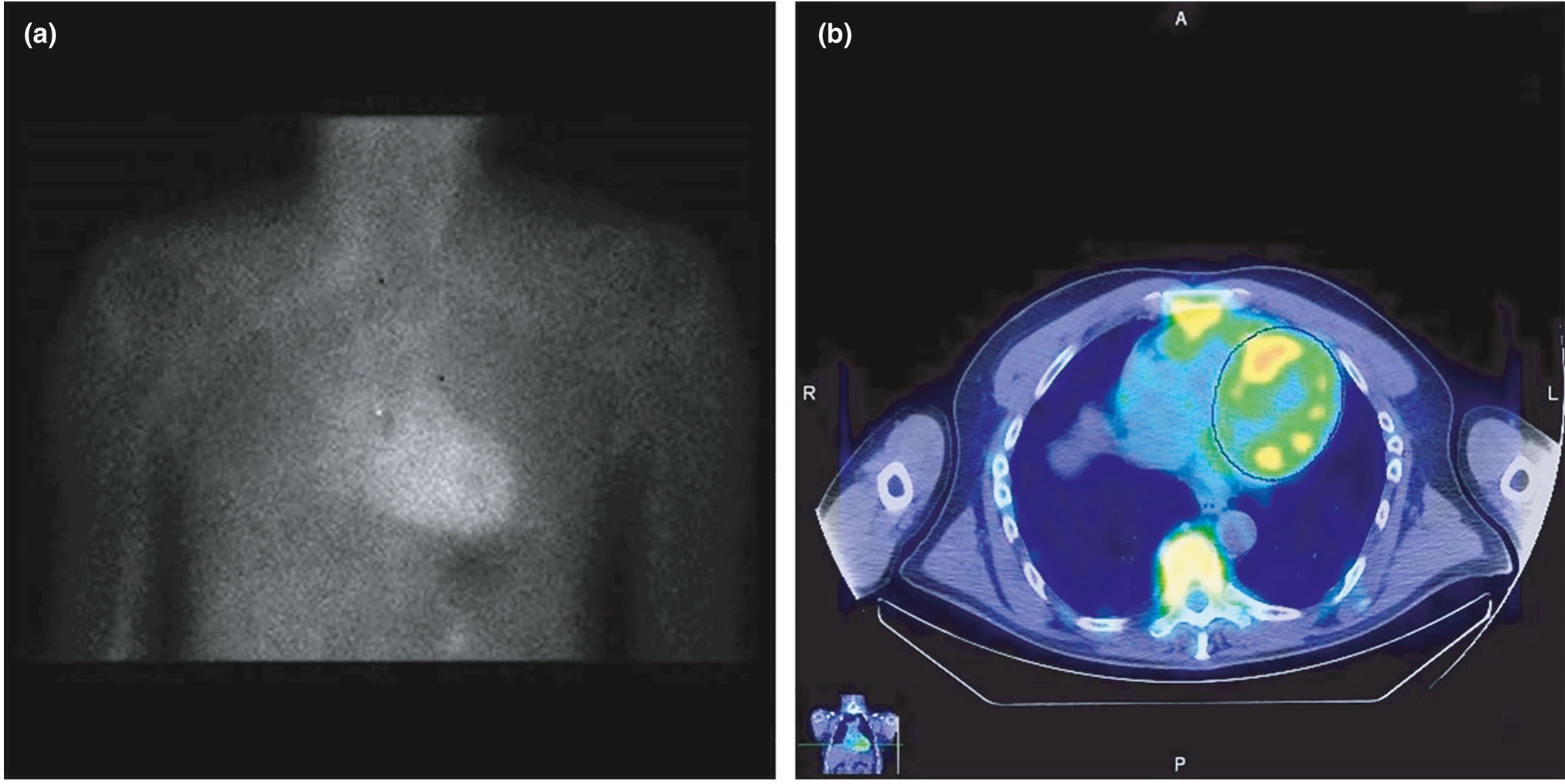
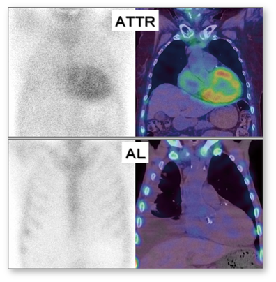
Notes: * The 99mTc phosphates currently in clinical use for cardiac amyloidosis diagnostics in Europe include 99mTc-DPD (3,3-diphosphono-1,2-propanodicarboxylate) and 99mTc-HMDP (hydroxymethylene-diphosphonate). By contrast, in the United States, 99mTc-PYP (pyrophosphate) is commonly used, while DPD is not approved. The exact mechanism that leads to the increased affinity of these radionuclides to amyloid deposits in the heart has not yet been clarified in detail but is suspected to be due to microcalcifications related to amyloid deposits. Also, the exact reason for the slightly higher affinity of these radionuclides for myocardial ATTR deposits compared to AL deposits is still unclear.2​

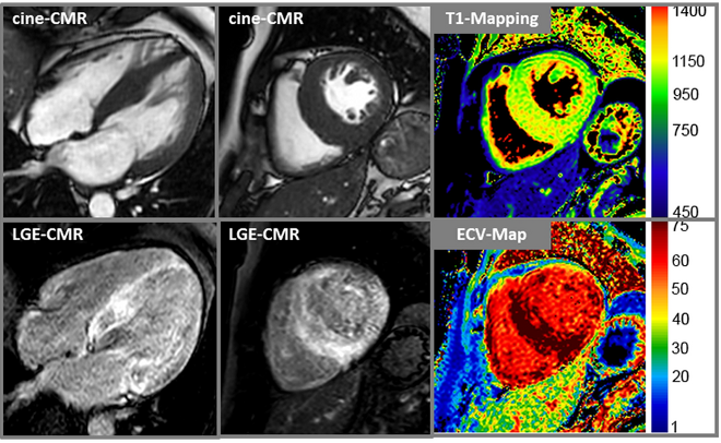
The main advantages of scintigraphy are its relatively low costs, its established reimbursement, the wider availability compared to CMR, and the independence of image quality from patient-specific factors. However, as outlined by Kittleson et al. recently, the test performance of scintigraphy in populations with lower disease prevalence is unknown and the aforementioned positive predictive value of scintigraphy (with the absence of monoclonal gammopathy) of 100% for ATTR is questioned by some recent reports that illustrate possible causes of false-positive 99mTc-PYP scans. In addition, first larger studies with a direct comparison of scintigraphic 99mTc-DPD images with a CMR-based determination of ECV in the same patients suggest that there is a prognostic significance for the more precise CMR-based ECV. Importantly, the decision whether to perform a multi-parametric CMR study and/or a 99mTc-phosphate scintigraphy for further non-invasive work-up of suspected cardiac amyloidosis depends (amongst others) on (a) local availability and (b) respective imaging expertise.2

  1. ALXN: ALXN2060 Differentiation workshop. Citing: Rapezzi, JACC Imaging. 2011:659-70 & Hutt, Eur Heart J. 2014;15:1289-1298.
  2. Yilmaz A, et al. Diagnosis and treatment of cardiac amyloidosis: position statement of the German Cardiac Society (DGK). Clin Res Cardiol. 110, 479–506 (2021). https://doi.org/10.1007/s00392-020-01799-3.
  3. Tahara N, et al. 99mTechnetium-pyrophosphate scintigraphy: a practical guide for early diagnosis of transthyretin amyloid cardiomyopathy. ESC Heart Failure. November 29, 2021. DOI:  https://doi.org/10.1002/ehf2.13693.

Native T1 Mapping for the Future of Diagnosis & Monitoring

CMR offers an indispensable method for the detection of CA, both in the context of classical methods (LGE) and by extension to the new mapping procedures, and should be used early in cases of unclear left-ventricular hypertrophy
In recent years, CMR has gained considerable importance in the diagnosis of amyloidosis and its distinction from other cardiomyopathies. The following 3 strengths are particularly important:
  1. The exact analysis of heart function and anatomy
  2. The recording of myocardial infiltration using contrast-enhanced images LGE:
    • A high-quality CMR study with a characteristic LGE finding is diagnostic of cardiac amyloidosis and similar to scintigraphy, the combination of such characteristic LGE findings with negative monoclonal protein studies will result in a high positive predictive value for the diagnosis of ATTR amyloidosis
  3. The recognition of diffuse changes in the myocardial extracellular space with specific mapping techniques:
    • With new methods, the magnetization of the heart muscle can be measured directly (“mapping”)
      • Mapping techniques do not require regional differences but show diffuse abnormalities by changing the absolute values
      • Native T1 mapping does not require a contrast agent and provides a quantitative measure of the severity of the disease
      • Native T1 mapping can detect cardiac amyloidosis as accurately as CM-assisted methods and shows significantly higher values for cardiac amyloidosis than in patients with hypertrophic cardiomyopathy
Responsive Image
In addition to the cine and LGE images, T1 mapping and ECV images are shown in the right column; particularly in the basal septum, measured values of T1 (native) = 1300 ms and ECV = 70% were significantly increased and characteristic for cardiac amyloidosis
While scintigraphic methods only allow for the diagnosis of amyloidosis, CMR offers the great advantage that different aetiologies associated with LV wall thickening or hypertrophy can be clarified simultaneously, and the exact underlying cause can usually be correctly assigned
  • Thus, the current HF guidelines of the European Society of Cardiology recommend a CMR study for tissue characterization in patients with HF symptoms (I/C)
  • Moreover, CMR is expected to play a central future role regarding non-invasive therapy monitoring in patients with cardiac amyloidosis receiving specific therapies due to its unique capabilities regarding non-invasive myocardial tissue characterization including depiction and quantification of amyloid load
Notes: CMR = cardiovascular magnetic resonance imaging; ECV = extracellular volume fraction; HF = heart failure; LGE = late gadolinium enhancement

Notes: * Patients with AL amyloidosis usually have more pronounced changes than patients with ATTR amyloidosis​

  • For both amyloid subtypes, the amount of native T1 time is closely linked to the prognosis​
  • The significance of possible differences between native T1 time, which indicates changes in intercellular and interstitial space, and ECV, which is determined by T1 mapping before and after CM administration and has a stronger emphasis on extracellular space, has not yet been conclusively clarified; however, the higher ECV values in ATTR compared to AL indicate pathophysiological differences at the cellular level between different types of amyloidosis ​
  • In a direct comparison of the hazard ratio values between ECV, LGE, and bone scintigraphy (based on the use of technetium [99mTc] phosphates), T1 and ECV data were prognostically superior to both LGE and scintigraphy ​
  • So far, however, these studies are relatively small monocentric studies with a rather short observation period.
  1. Yilmaz, A., Bauersachs, J., Bengel, F. et al. Diagnosis and treatment of cardiac amyloidosis: position statement of the German Cardiac Society (DGK). Clin Res Cardiol 110, 479–506 (2021). https://doi.org/10.1007/s00392-020-01799-3

Where / What to Biopsy?

In the pathological diagnosis of CA, the detection rate of amyloid deposits by myocardial biopsy is almost 100%, and is considered the gold standard for diagnosing this disorder [1]
Responsive Image
However, in some cases, especially in older patients, performing a myocardial biopsy involves an increased risk for complications
  • In CA, it is necessary to distinguish between the three typical types of amyloidosis, i.e., AL, ATTRwt, and ATTRv, and to introduce treatment specific to each disease at an early stage
  • To establish the diagnosis of AL amyloidosis, it is important to confirm the presence of amyloid deposits derived from immunoglobulin light chain in any organ
  • On the other hand, in the early stages of ATTRwt, amyloid deposits are difficult to detect in biopsies at sites other than the heart; thus, clinical diagnostic methods using diagnostic imaging such as PYP scintigraphy have been proposed in the EU and US [1]
Detection Rate of Amyloid at Screening Biopsy Sites in CA
Responsive Image
In the case of ATTRwt, the detection rate is low for amyloid in abdominal fat pad needle aspiration biopsy, skin biopsy, gastrointestinal biopsy, lip biopsy, etc.; thus, if amyloid is not detected at these biopsy sites, a myocardial biopsy should be planned [1]
japan Responsive Image
Per the Japanese Guidelines: when combined with other diagnostic tests, detection of amyloid fibrils in biopsies obtained from organs other than the heart through less invasive procedures may be used to confirm diagnosis, especially when obtaining a cardiac biopsy may be difficult [1]
* Note: a recent (2021) study of 78 ATTRwt patients suggests that females have a higher sensitivity to TTR deposition from abdominal fat than male patients

** When performing deeper and wider biopsies than usual, e.g., a study of 11 ATTRwt patients (6 males and 5 females) who underwent surgical skin biopsy from the abdominal wall showed 73% sensitivity; deposits were seen mainly in the deep layer of subcutaneous fat tissue and showed a patchy distribution [1,4]

*** Examination in a few cases (n = 3/8) [5]
Deep Dive
Gender differences in biopsy predictiveness
Go to Deep Dive
ALXN: 32_2021 Amyloidosis March KOL Perspectives_Cristina Quarta_FINAL. Citing:
  1. Kitaoka H, et al. Circ J. 2020;84:1610-1671.
  2. Muchtar E, et al. J Intern Med. 2021;289:268-292.
  3. Veinot JP. Can J Cardiol. 2009;25:e55-e56.
  4. Ikeda S, Sekijima Y, Tojo K, Koyama J. Diagnostic value of abdominal wall fat pad biopsy in senile systemic amyloidosis. Amyloid. 2011 Dec;18(4):211-5. doi: 10.3109/13506129.2011.623199. Epub 2011 Oct 17. PMID: 22004460.
  5. Ueda M, Horibata Y, Shono M, Misumi Y, Oshima T, Su Y, Tasaki M, Shinriki S, Kawahara S, Jono H, Obayashi K, Ogawa H, Ando Y. Clinicopathological features of senile systemic amyloidosis: an ante- and post-mortem study. Mod Pathol. 2011 Dec;24(12):1533-44. doi: 10.1038/modpathol.2011.117. Epub 2011 Aug 5. PMID: 21822203.

Staging ATTR Amyloidosis

Currently, there is no universal system of staging ATTR amyloidosis, but biomarker-based staging systems have been developed by the Mayo Clinic and the NAC [1,6]
Responsive Image
  • Mayo Clinic model: soluble cardiac biomarkers are able to stratify ATTRwt patients into three distinct risk groups based on troponin T and NT-proBNP
    • Patients who do not meet thresholds, meet one threshold, or meet both have 4-year survival rates of 57%, 42% and 18%, respectively
    • The median survival of ATTRwt in a large Mayo cohort was 3.6 years (n=360 ATTRwt patients)
  • NAC (UK) model: replaces troponin T with eGFR, while keeping NT-proBNP at the same Mayo Clinic model threshold
    • The NAC criteria are based on biomarkers of cardiomyopathy (NT-proBNP) and nephropathy (eGFR) and can determine disease severity
      • Reflect the cardiomyopathy predominantly associated with ATTRwt amyloidosis and, to a lesser extent, with ATTRv amyloidosis
      • Reflect the renal comorbidity frequently occurring in the elderly [2,3]
There is a lack of a staging system that includes criteria for peripheral or autonomic polyneuropathy [1]


Notes: BNP = brain natriuretic peptide; NAC = National Amyloidosis Centre (UK); NT-proBNP = N-terminal pro b-type natriuretic peptide; eGFR = estimated glomerular filtration rate
Deep Dive
Mayo Model and NAC Model 
Go to Deep Dive
Deep Dive
Prognosis & NYHA Class
Go to Deep Dive
  • The NYHA classification is not currently used [4,5]
    • However, there is a correlation between the NYHA class at diagnosis and treatment response4
  • NYHA class ≥III is associated with poorer survival [5]
  1. ALXN: Acoramidis ​for the Treatment of ATTR-CM: Training Deck. November 22, 2021. 34_Acoradmidis Training Deck_22Nov21 FINAL. Citing: Gillmore JD, Damy T, Fontana M, Hutchinson M, Lachmann HJ, Martinez-Naharro A, et al. A new staging system for cardiac transthyretin amyloidosis. Eur Heart J. 2018;39(30):2799-806.
  2. ALXN: ATTR Amyloidosis: Epidemiology Dossier. Elissa Wilker & Shona Fang. September 2021. 18_ATTR Amyloidosis Epi Dossier. Citing: Grogan M, Scott CG, Kyle RA, Zeldenrust SR, Gertz MA, Lin G, et al. Natural History of Wild-Type Transthyretin Cardiac Amyloidosis and Risk Stratification Using a Novel Staging System. J Am Coll Cardiol. 2016;68(10):1014-20 & Gillmore JD, et al. Eur Heart J. 2018;39:2799-2806.
  3. Muchtar E, et al. Systemic amyloidosis from A (AA) to T (ATTR): a review. J Intern Med. 2021;289(3), 268-292. https://doi.org/10.1111/joim.13169. Hazard ratios for the ATTR Mayo model and National Amyloidosis Centre model are derived from the original publications.
  4. ALXN: Acoramidis ​for the Treatment of ATTR-CM: Training Deck. November 22, 2021. 34_Acoradmidis Training Deck_22Nov21 FINAL. Citing: Kittleson MM, et al. Circulation. 2020;142:e7-e22. Erratum in: Circulation. 2021;144:e11.
  5. ALXN: Acoramidis ​for the Treatment of ATTR-CM: Training Deck. November 22, 2021. 34_Acoradmidis Training Deck_22Nov21 FINAL. Citing: Sperry BW, et al. J Am Heart Assoc. 2016;5:e002877.
  6. Garcia-Pavia P, et al. Diagnosis and Treatment of Cardiac Amyloidosis: A Position Statement of the ESC Working Group on Myocardial and Pericardial Diseases. Eur Heart J. 2021;42(16):1554-1568. DOI: https://doi.org/10.1093/eurheartj/ehab072.