Diagnosing Physicians

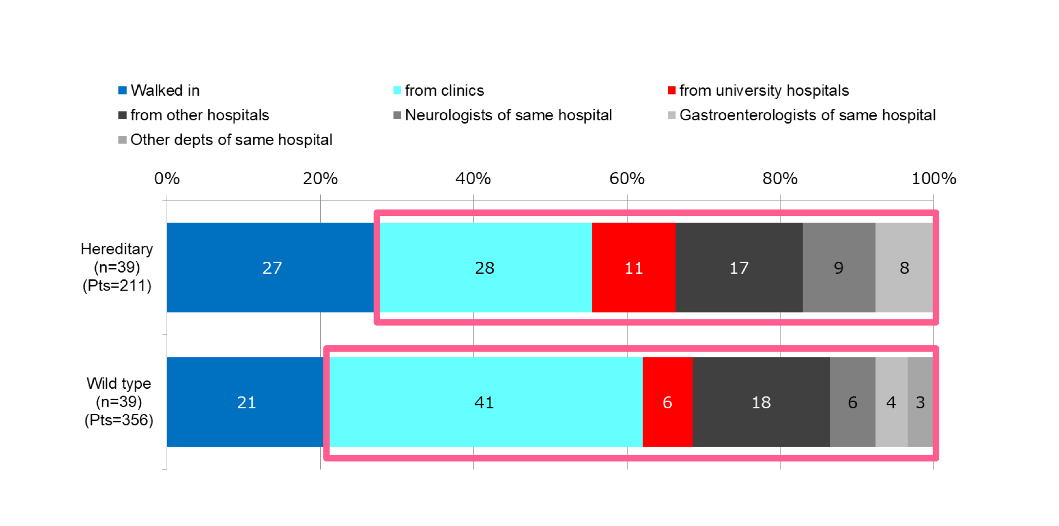
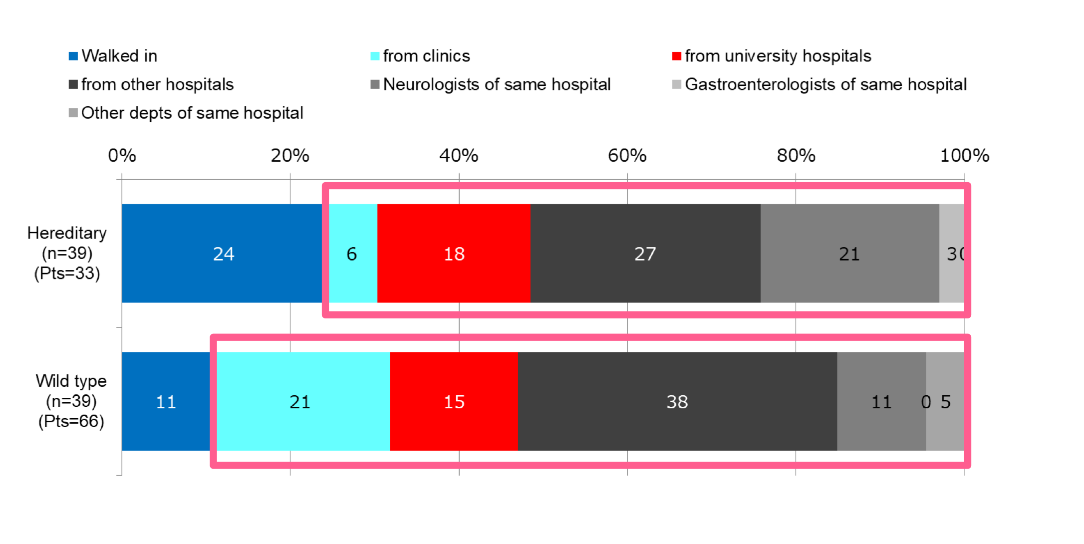
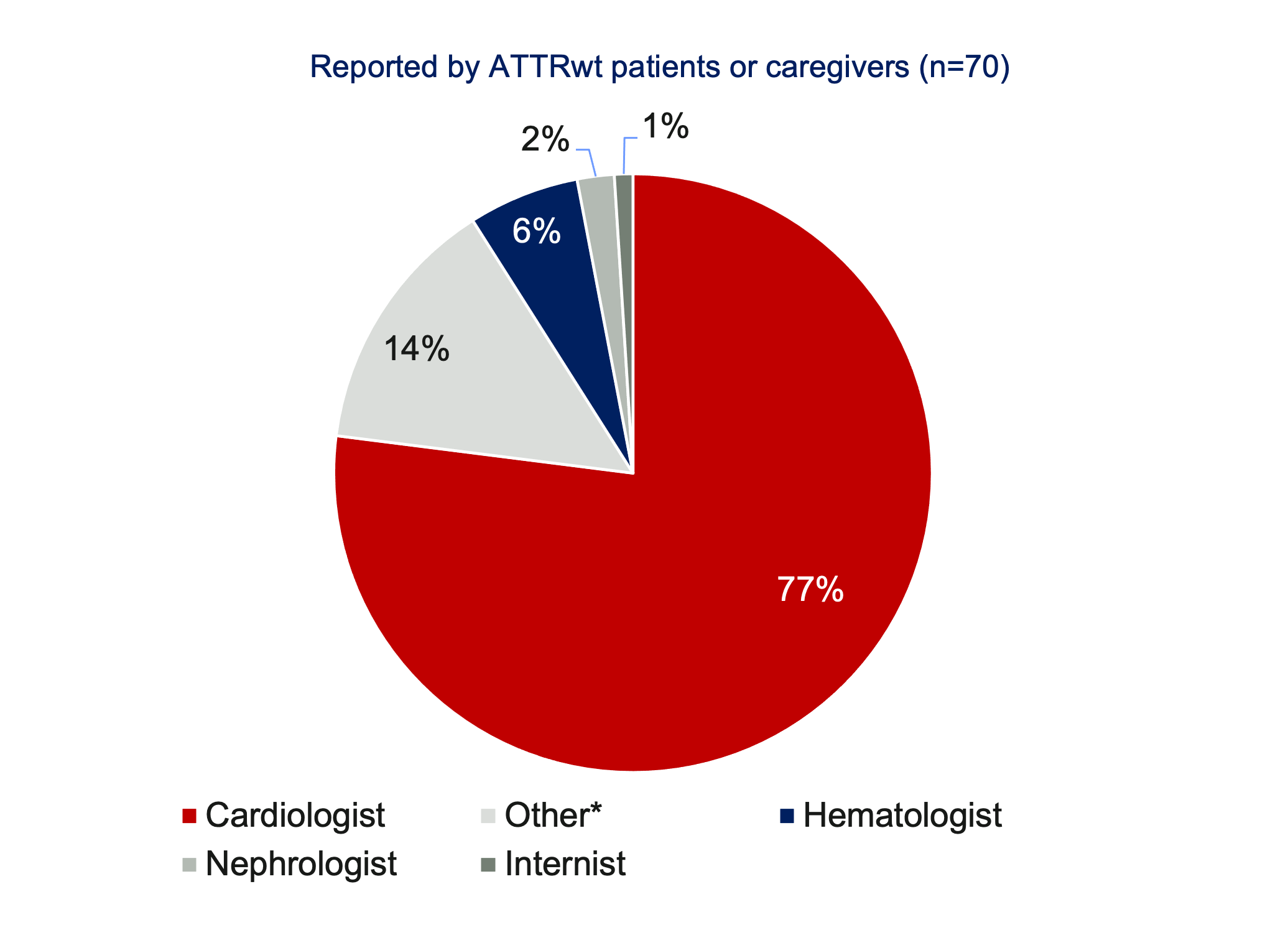
Many specialties are involved in the diagnosis of ATTR; but cardiologists are most often the diagnosing physician for ATTRwt [1,2]
SPECIALTY OF DIAGNOSING PHYSICIAN [1]
All Types of Amyloidosis
Responsive Image
GI = Gastroenterologist; HEME/ONC = Hematologist / Oncologist
SPECIALTY OF DIAGNOSING PHYSICIAN [2]**
ATTRwt Amyloidosis
Responsive Image
Amyloidosis Survey: Results from Patients with AL and ATTR Amyloidosis and Their Caregivers. Presented at the XVIth International Symposium on Amyloidosis. 2018. Kumamoto, Japan.​

Notes: Other includes internists, hepatologists, ophthalmologists & dermatologists​

Notes: Surveys created by the ARC were made available on the ARC website and were given to the Amyloidosis Foundation, Amyloidosis Support Groups, and individual physicians for distribution. Of the overall total (n=699 all respondents), 54% were from the Asia-Pacific region, 17% from the EU, and 75% from North America. 

  1. Lousada, et al. Light Chain Amyloidosis: Patient Experience Survey from the Amyloidosis Research Consortium. Adv. Ther. (2015) 32:920–28. DOI: 10.1007/s12325-015-0250-0.
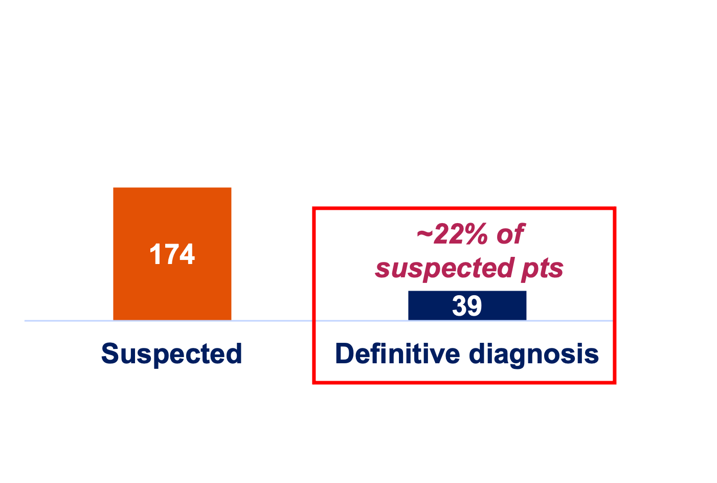
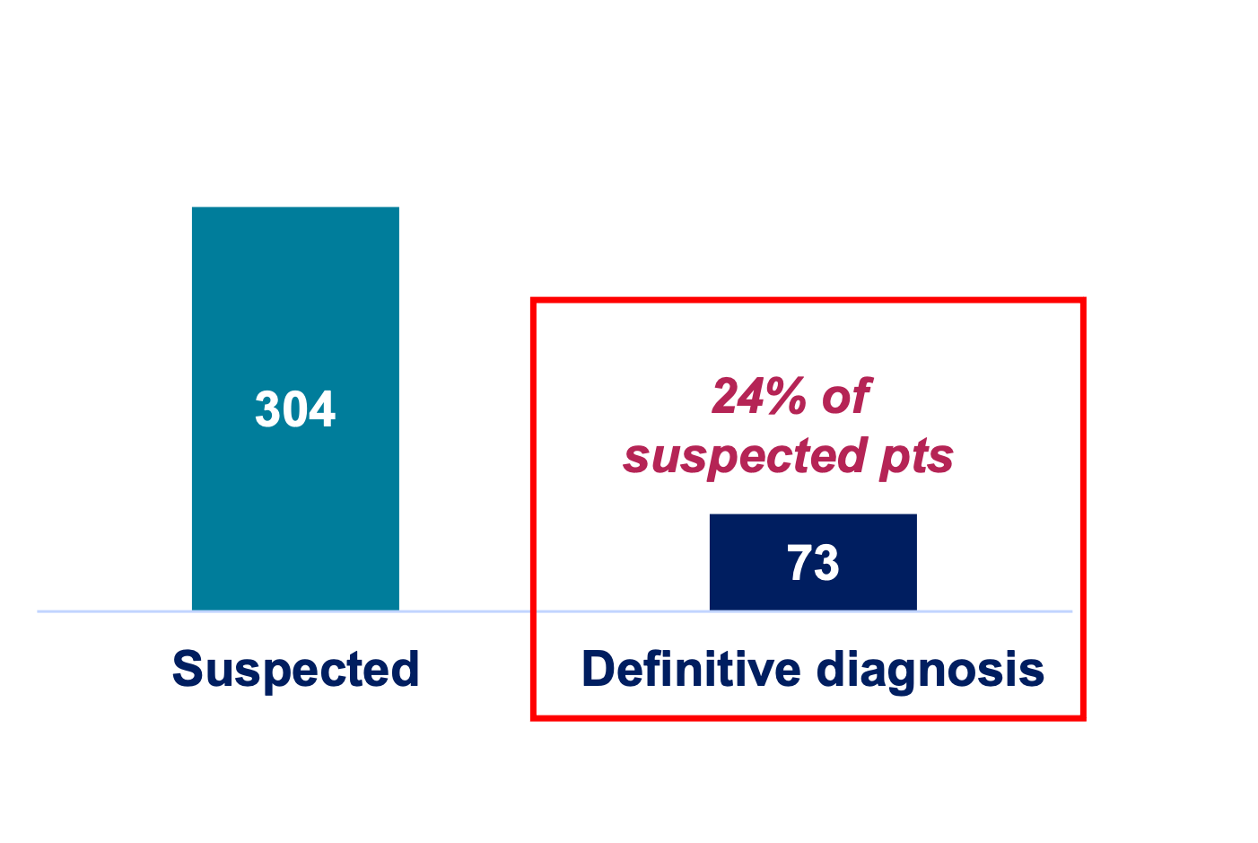
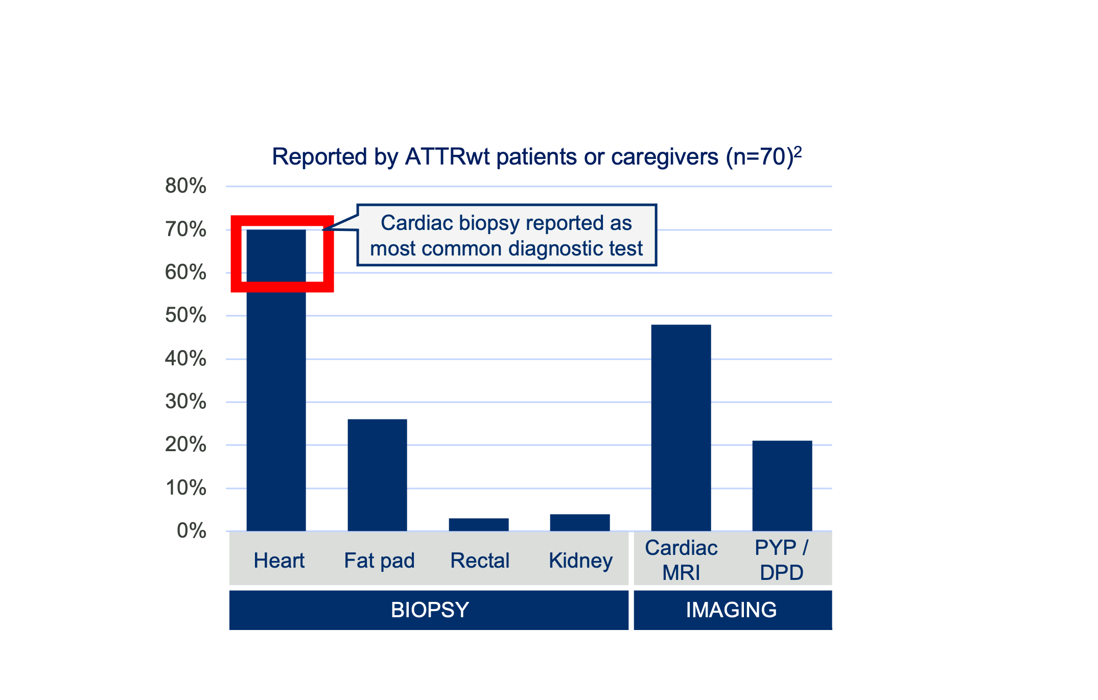
    • Table 2 Establishment of amyloidosis diagnosis
  2. Lousada, et al. Amyloidosis Research Consortium Cardiac Amyloidosis Survey: Results from Patients with AL and ATTR Amyloidosis and Their Caregivers. Presented at the XVIth International Symposium on Amyloidosis ISA (2018) in Kumamoto.

Quantitative Survey

Japan Responsive Image
Quantitative Survey: Patients Suspected of vs. Diagnosed with ATTR-CM
Less than a quarter of patients reached a definitive ATTR diagnosis at institutions that are unapproved for use of the sole disease-modifying therapy currently available in Japan for ATTRwt
Vyndaqel prescribers
< Past 1 year >
At institutions where Vyndaqel is approved, > ½ eventually reached a definitive diagnosis
Responsive Image
Hereditary ATTR-CM (# of patients)
Responsive Image
Wild type ATTR-CM (# of patients)
Responsive Image
Drs. of institutions not approved for Vyndaqel
< Past 1 year >
At institutions where Vyndaqel is NOT approved, ~ ¼ reached a definitive diagnosis
Ratios of using myocardial biopsy and pyrophosphate scintigraphy were lower among diagnostic tests performed
Responsive Image
Hereditary ATTR-CM (# of patients)
Responsive Image
Wild type ATTR-CM (# of patients)
Responsive Image
Copyrightⓒ INTAGE Healthcare Inc. All Rights Reserved.
Referral Patterns
The majority of ATTRwt-CM patients (79% of suspected patients and 89% of those with a definitive diagnosis) came in as referrals
Suspected Patients
How suspected patients present:
  • Of the patients suspected of ATTR-CM that were seen at institutions, 73% of hereditary and 79% of wild types came in as referrals
  • Many were from clinics which shared, around 30% and 40% for hereditary and wild type, respectively
Responsive Image
Patients already with definitive diagnosis
How patients already with definitive diagnosis present:
  • Of the patients who were already diagnosed with ATTR-CM that were seen at institutions, 76% of hereditary and 89% of wild types came in as referrals
  • Many were from “hospitals other than universities” which shared around 30-40%
Responsive Image
Copyrightⓒ INTAGE Healthcare Inc. All Rights Reserved.
  1. ALXN: Acceptance of ALXN2060 for ATTR-CM (Quantitative Survey). October 28, 2021. 02_210823_ATTR-CM quantitative research.

Suspecting Amyloidosis in General (Any Type)

When to Suspect Amyloidosis
Responsive Image
The different forms of amyloidosis may impact any tissue and organ system, with a spectrum of clinical manifestations—these in turn can coexist both within a single patient and between different types of amyloidosis
Early diagnosis relies upon a high index of suspicion, and the following features, particularly the coexistence of ≥2 clinical features and/or clinical features in the context of predisposing conditions:

"Red flag" clinical features:
  • Non-organ specific: unintentional weight loss, loss of appetite, severe fatigue
  • Visible tissue infiltration: macroglossia, easy bruising, skin fragility, nail dystrophy, waxy or thickened skin
  • Proteinuria with or without nephrotic syndrome
  • Heart failure
  • Orthostatic hypotension, arrhythmia
  • Carpal tunnel syndrome and/or progressive "glove and stocking" peripheral neuropathy
  • Features of autonomic dysfunction including constipation and/or diarrhea, orthostatic hypotension, erectile dysfunction, gustatory sweating
  • Hepatosplenomegaly
Known underlying predisposing conditions:
  • Monoclonal gammopathy, multiple myeloma, or other lymphoplasmacytic disorders known to result in production of monoclonal immunoglobulins
  • Persistent uncontrolled inflammatory diseases (e.g., rheumatoid arthritis (RA), inflammatory bowel disease), or chronic infection (e.g., bronchiectasis, urosepsis)
  • A family history of amyloidosis, neuropathy, renal disease, cardiomyopathy, or of inflammatory symptoms, suggesting inherited
Diagnosis is usually a complicated process . . .
  • The definitive diagnostic method is tissue biopsy, although presence of amyloidosis may be suggested by the history and clinical manifestations (e.g., nephrotic syndrome in a patient with multiple myeloma or longstanding, active RA) 
  • In many patients, the biopsy need not be from the known affected organ but can be from another site likely to have deposits, e.g., bone marrow or abdominal fat pad
  • In some patients, the presence of amyloid is demonstrated by findings on imaging
  • In some patients, a biopsy result consistent with amyloid is an unexpected diagnosis following routine laboratory Congo Red staining (e.g., AA amyloidosis is only one cause of the nephrotic syndrome in RA patients)
  • Even when amyloidosis is expected, tissue biopsy is important because assumptions regarding the type of amyloid may be incorrect
Deep Dive
Typical clinical and lab findings
Go to Deep Dive

When to Suspect ATTR-CM

ATTR-CM amyloidosis should be suspected if there are multiple nonspecific symptoms [1,2]
Responsive Image

Glossary:

  • Angina, a type of chest pain or discomfort caused by reduced blood supply to the heart​
  • Angiogram, an X-ray imaging technique that helps evaluate blocks in blood vessels and organs​
  • Pericardial effusion, the buildup of excess fluid in the sac-like structure surrounding the heart (pericardium) ​
  • Pleural effusions, the buildup of excess fluid between the layers lining the lungs and the inside of the chest cavity (pleura)​
  • Pseudo-infarction, any condition that mimics a heart attack
ALXN: Acoramidis ​for the Treatment of ATTR-CM: Training Deck. November 22, 2021. 34_Acoradmidis Training Deck_22Nov21 FINAL. Citing:
  1. Yamamato H, Yokochi T. ESC Heart Fail. 2019;6:1128-1139.
  2. Kittleson MM, et al. Circulation. 2020;142:e7-e22. Erratum in: Circulation. 2021;144:e11.

Suspecting and Diagnosing ATTRwt-CM

Several research groups have discussed the early signs and symptoms (red-flag symptom clusters) of ATTRwt-CM and proposed diagnostic algorithms; (unexplained) LVH features prominently [1]
Responsive Image
Illustration of a sequential algorithm to ensure early diagnosis [1]:
A suspected diagnosis of ATTRwt‐CM should be triggered by unexplained LVH plus at least one of the following:
  • Presence of HF symptoms
  • Elevated laboratory test results (NT‐proBNP, hs‐TnT, or hs‐TnI)
  • Abnormal ECG findings
  • Imaging (ECHO or CMR) findings characteristic of ATTRwt‐CM
  • Advanced age (≥60 years)
  • Medical history suggestive of ATTRwt‐CM (CTS, spinal canal stenosis)

If a patient with unexplained LVH meets ≥1 criterion listed above, a detailed examination using 99mTc‐PYP bone scintigraphy and monoclonal protein detection tests should be offered to exclude AL amyloidosis and grade the cardiac 99mTc‐PYP uptake

To differentiate between ATTRv‐CM and ATTRwt‐CM, tissue biopsy, amyloid typing, and TTR genetic testing should be considered
Responsive Image
A medical advisory board meeting was held in Tokyo, Japan, on 3 February 2019. This diagnostic algorithm was developed based on discussions held at this meeting.

Notes: CMR = cardiac magnetic resonance; CTS = carpal tunnel syndrome; ECG = electrocardiogram; ECHO = echocardiogram; FLC = free light chain; hs‐TnI = high‐sensitivity troponin I; hs‐TnT = high‐sensitivity troponin T; LVH = left ventricular hypertrophy; NT‐proBNP = N‐terminal pro‐B‐type natriuretic peptide; 99mTc‐PYP = 99mTechnetium‐pyrophosphate
 
Deep Dive
Cardiac & extracardiac red flags
Go to Deep Dive
When to Suspect ATTRwt-CM During Routine Cardiac Tests
Japan Responsive Image
Responsive Image
The diagnosis may also be suspected because of typical symptoms and/or the results of a routine cardiac test—an electrocardiogram or echocardiogram
Barriers to “suspecting”
Another form of cardiac amyloidosis, known as light-chain amyloidosis (AL), must first be ruled out, as AL and ATTR-CM are managed in different ways
  • AL amyloidosis may be ruled out using blood and urine tests*
  • Endocardial biopsy currently also remains in use as a method to distinguish between AL and ATTR-CM [3]

Once suspected, more specialized tests are needed to confirm the diagnosis; these could include:
  • Imaging studies of the heart, most commonly a cardiac MRI and/or a nuclear medicine scan of the heart
  • A tissue biopsy of an affected organ
  • Genetic testing (to rule out ATTRv) [2]

In consideration of Japanese daily medical practice, cardiac pump dysfunction (HF), abnormal heart rhythms (arrhythmia), investigation of cardiac involvement in patients already diagnosed as having amyloidosis in other organs, and cardiac findings during medical check-ups could serve to highlight a suspected diagnosis of ATTRwt-CM [1]

Generally, in Japan, patients presenting with cardiac symptoms undergo an echocardiogram as an initial step, regardless of HF symptoms; thus, it is advisable to carefully examine echocardiographic findings for warning signs such as unexplained left ventricular hypertrophy (LVH) [1]
 
Diagnostic Procedures Performed
ATTRwt Patients with Cardiac Amyloidosis (CA)
Responsive Image
Presented at the XVIth International Symposium on Amyloidosis (2018) in Kumamoto, Japan
Endocardial biopsy has been the gold standard for diagnosing ATTR-CM because of its high sensitivity and specificity—it is currently a standard for diagnosis through the JCS guidelines but may become less common due to advances in imaging [4]
Notes: JCS guidelines = Japan Cardiology Society Guideline on Diagnosis and Treatment of Cardiac Amyloidosis
Notes: *E.g., tests for abnormal antibody (immunoglobulin) proteins in the blood:  ​
  • Freelite Assay (or “FLC” for free light chains), which shows the level of kappa and lambda light chains​
  • Immunofixation electrophoresis, which can be done with blood and/or urine5
  1. American Heart Association. What is Transthyretin Amyloid Cardiomyopathy (ATTR-CM)? https://www.heart.org/-/media/files/health-topics/answers-by-heart/what-is-attrcm.pdf?la=en.
  2. Inomata, T, et al. Diagnosis of wild-type transthyretin amyloid cardiomyopathy in Japan: red-flag symptom clusters and diagnostic algorithm. ESC Heart Failure. 2021. DOI: 10.1002/ehf2.13473.
  3. Hafeez AS, Bavry AA. Diagnosis of Transthyretin Amyloid Cardiomyopathy. Cardiol Ther. 2020;9(1):85-95.
  4. Kitaoka H, et al. JCS 2020 guideline on diagnosis and treatment of cardiac amyloidosis. Circ J. 2020. 84(9), 1610-1671. https://doi.org/10.1253/circj.CJ-20-0110.
  5. https://www.yourheartsmessage.com/attr-cm-diagnosis.